Reconstruction
Magnetic resonance (MR) imaging can provide detailed anatomical information during interventions. However, the raw data obtained from MR scans often require sophisticated processing techniques for accurate and meaningful interpretation. This is where the concept of reconstruction comes into play. MR image reconstruction involves the conversion of acquired raw data into a visual representation that accurately reflects the internal structures of the imaged anatomy. This process employs advanced mathematical algorithms and computational methods to enhance image quality, reduce artifacts, and improve diagnostic accuracy:
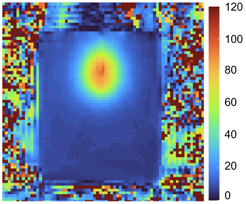
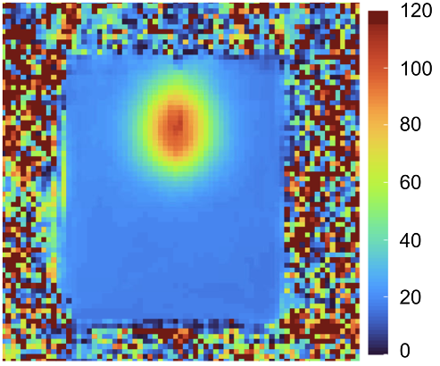
Accurate and reliable temperature measurements are essential during thermometry based on the proton resonance frequency shift (PRFS), and phase drift correction is crucial. Given the high temporal resolution required for PRFS-thermometry, the MR scanner’s hardware is under significant strain, particularly its gradient system. Even minor changes in the main magnetic field can substantially impact the phase images obtained. However, with the help of phase drift correction algorithms [1], these minor fluctuations in the magnetic field are accounted for, allowing for precise phase images to be obtained with confidence. This correction is especially crucial in hyperthermia and thermal ablative interventions, where accurate temperature knowledge of the targeted area is essential for optimal clinical outcomes.
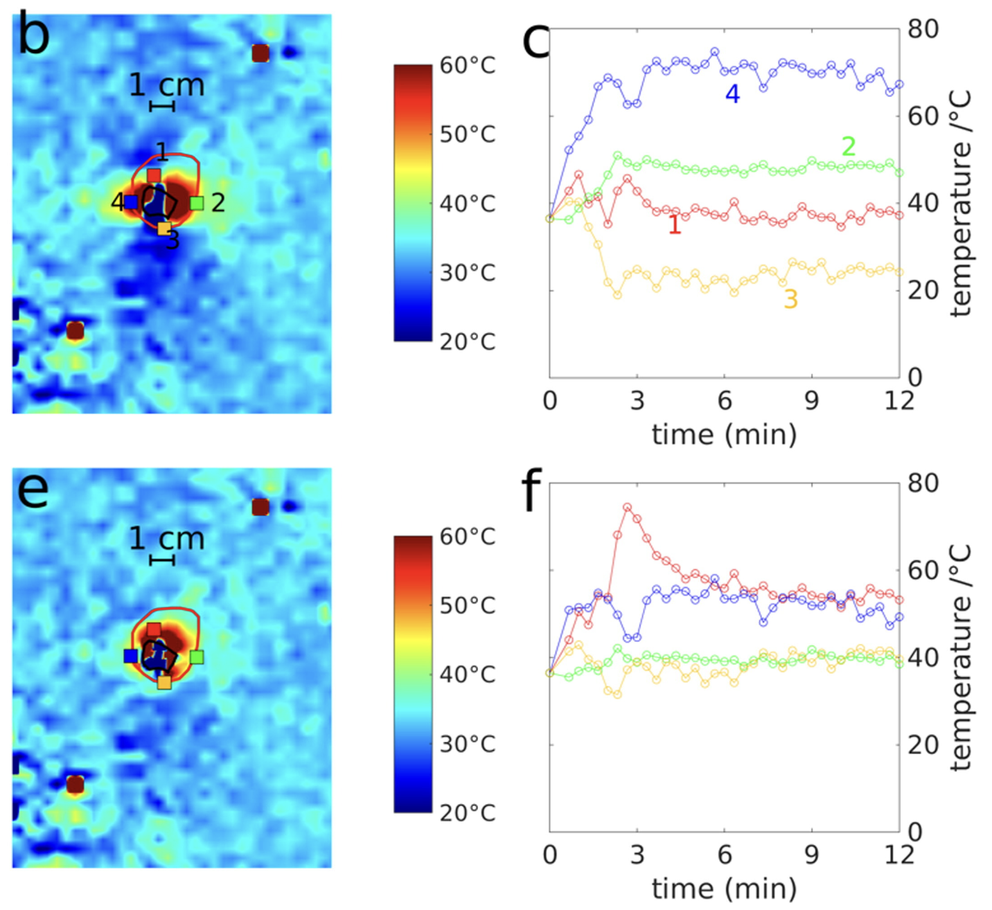
Temperature maps are a reliable tool for estimating the damage caused to tumors by thermal ablation procedures. When performing microwave ablation, high temperatures are generated in the vicinity of the applicator, which can result in tissue carbonization and gas formation. These changes cause magnetic susceptibility artifacts in the target area. However, an artifact correction algorithm can account for these distortions, allowing for more precise temperature estimations [2]. This is particularly crucial in MR-guided thermal ablations involving high target temperatures, as it ensures dependable and accurate outcomes.
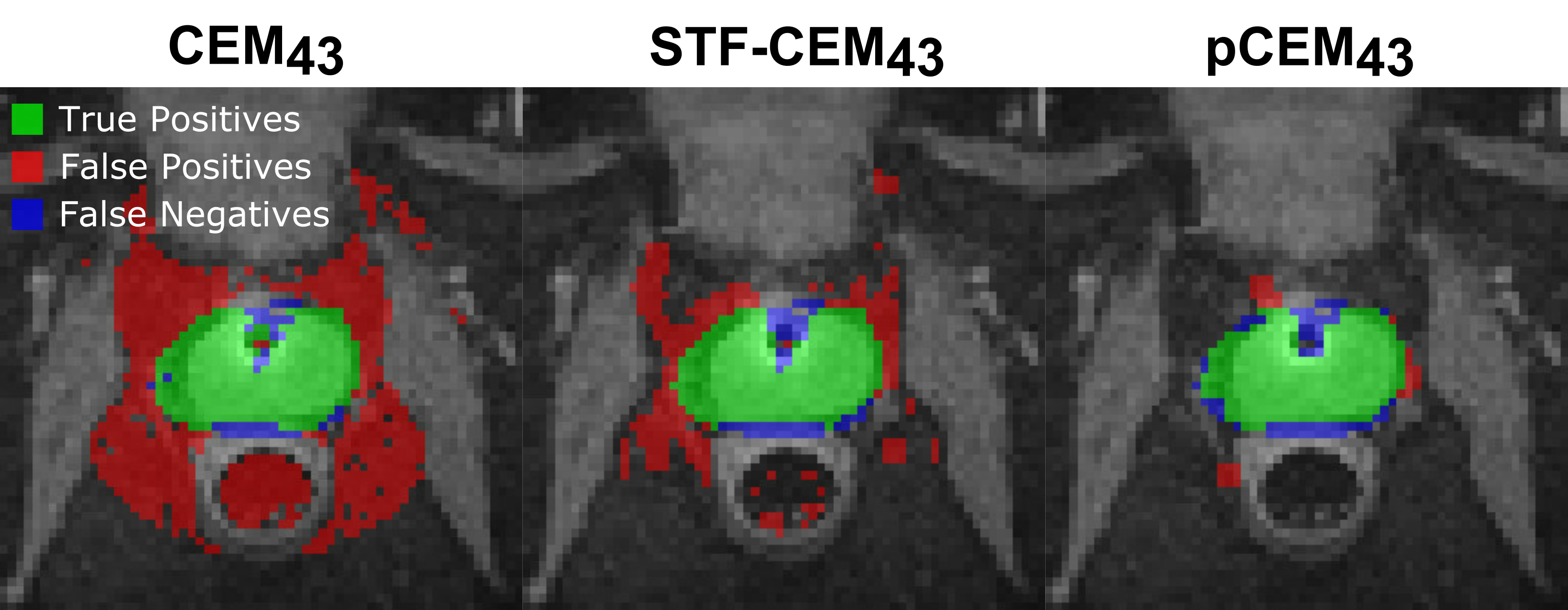
Thermal dose models such as the pCEM43 model [3][4] offer a range of benefits that enhance the precision and effectiveness of thermal treatments. These models provide a comprehensive and quantitative assessment of the thermal energy delivered to tissues over time, taking into account factors like temperature, exposure duration, and tissue characteristics. By integrating these models with MR thermometry data, clinicians can accurately predict the cumulative thermal effect on tissues, aiding in treatment planning and real-time monitoring. These models enable clinicians to tailor treatment parameters, optimize thermal doses, and ensure that therapeutic thresholds are achieved while minimizing the risk of undertreatment or overtreatment.
The pCEM43 model [3][4] is an improvement on the commonly used CEM43 model [5] that takes into account additional information about the noise statistics of the measurements allowing more accurate estimation of necrotic tissue during ablation procedures.
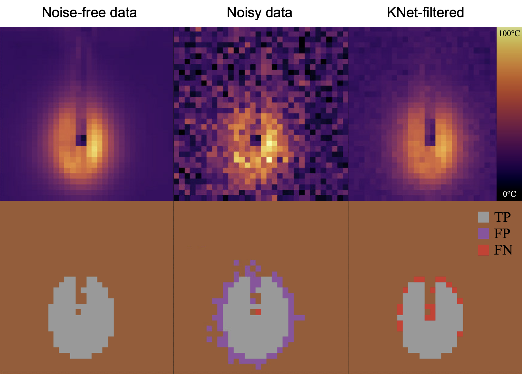
KalmanNet, an innovative approach that combines Kalman filtering and neural networks [6], offers compelling benefits in the field of MR thermometry. By leveraging the predictive power of neural networks and the state estimation capabilities of Kalman filters, KalmanNet enhances the accuracy and robustness of temperature mapping in real-time MRI [7]. Its ability to effectively predict and correct for noise and artifacts in MR thermometry data enables clinicians to obtain more reliable and consistent temperature measurements, even in challenging imaging conditions.
References
[1] Belker et al. Evaluating Various Phase Drift Correction Methods in PRFS-based Thermometry in the Pelvic Region of Free-Breathing Volunteers. ISMRM Proceedings (2023).
[2] Hensen et al. Correction of heat-induced susceptibility changes in respiratory-triggered 2D-PRF-based thermometry for monitoring of magnetic resonance-guided hepatic microwave ablation in a human-like in vivo porcine model. International journal of hyperthermia 39 1 (2022): 1387-1396.
[3] Schröer et al. A probabilistic thermal dose model for the estimation of necrosis in MR-guided tumor ablations. Medical Physics 51 1 (2023).
[4] Schröer et al. Improving PRFS-based MR Thermometry in Prediction of Ablation Zones during MR-guided Prostate Ultrasound Ablation using a Probabilistic Thermal Dose Model. ESMRMB Proceedings (2023).
[5] W C. Dewey. Arrhenius relationships from the molecule and cell to the clinic. International journal of hyperthermia 25 1 (2009): 3-20.
[6] G. Revach et al. KalmanNet: Neural Network Aided Kalman Filtering for Partially Known Dynamics. IEEE Transactions on Signal Processing 70 (2022): 1532-1547.
[7] Schröer et al. KalmanNet in MR-Thermometry: A step towards accurate real-time 3D monitoring of thermoablation procedures. RSNA Proceedings (2022).